
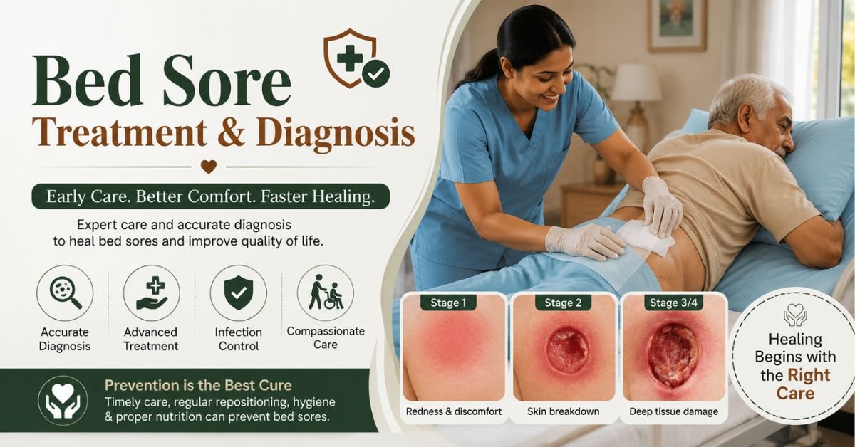
Getting the right bed sore treatment support starts with an accurate bed sore diagnosis — without one, even good care can miss the mark. Pressure ulcers form when skin sits against a bed, chair, or medical device too long, cutting off blood flow, and a correct bed sore diagnosis determines which bed sore treatment support plan will actually work. Left unchecked, a mild red patch can progress into an open wound reaching muscle or bone within days. This guide covers every stage, how clinicians diagnose bed sores, evidence-based treatment options, home-care strategies, and when to seek emergency help — based on current clinical guidance.
What Is a Bed Sore? (Quick Overview)
A bed sore — also called a pressure ulcer, pressure injury, or decubitus ulcer — is localized damage to skin and underlying tissue, usually over a bony area like the tailbone, heels, hips, or elbows. They’re caused by sustained pressure, often combined with friction or shear force against a surface, as described in detail by the Cleveland Clinic.
Anyone with limited mobility is at risk, including people who are bedridden, use a wheelchair, or have reduced sensation from conditions like spinal cord injury or diabetic neuropathy. Experts estimate roughly 2.5 million Americans develop bed sores every year, and worldwide, bed sores contribute to more than 24,000 deaths annually when infections go untreated.
Causes and Risk Factors
Bed sores don’t appear randomly — they form when specific risk factors combine. Understanding these helps both diagnosis and prevention.
Primary Causes
- Sustained pressure that compresses blood vessels and cuts off oxygen to tissue
- Friction from skin rubbing against sheets or clothing during repositioning
- Shear force when skin moves in one direction while underlying bone moves in another (common when a bed’s head is raised and the body slides down)
- Moisture from sweat, urine, or wound drainage that softens and weakens skin
Who Is Most at Risk
| Risk Factor | Why It Increases Risk |
|---|---|
| Limited mobility (bed rest, wheelchair use, paralysis) | Cannot independently shift weight off pressure points |
| Reduced sensory perception (spinal cord injury, neuropathy) | Can’t feel discomfort that would normally prompt movement |
| Poor nutrition or low body weight | Less natural cushioning and slower tissue repair |
| Incontinence | Chronic moisture weakens skin’s protective barrier |
| Advanced age | Skin becomes thinner and less elastic over time |
| Chronic conditions (diabetes, vascular disease) | Impaired blood flow slows healing and increases vulnerability |
| Cognitive impairment (dementia) | May be unable to communicate discomfort or reposition independently |
Bed Sore Stages: How Diagnosis Begins
Correct staging is the foundation of any bed sore diagnosis, because treatment differs significantly by depth of tissue damage. The National Pressure Injury Advisory Panel (NPIAP) staging system is the clinical standard used across hospitals and home health settings, and is also detailed in the StatPearls clinical reference.
Bed Sore Stages at a Glance
| Stage | What It Looks Like | Tissue Depth | Typical Healing Time |
|---|---|---|---|
| Stage 1 | Red area that doesn’t turn white (blanch) when pressed; skin intact | Top skin layer only | Days, if caught early |
| Stage 2 | Shallow open wound or blister; pink/red wound bed | Epidermis + partial dermis | 1–4 weeks |
| Stage 3 | Crater-like wound; fat may be visible | Full-thickness skin loss | 1–4 months |
| Stage 4 | Deep wound exposing muscle, tendon, or bone | Full-thickness + deeper structures | 3 months–2 years |
| Unstageable | Wound base covered by dead tissue (slough/eschar), true depth hidden | Unknown until debrided | Varies |
| Deep Tissue Injury | Purple/maroon intact or blood-filled area; damage below the surface | Underlying soft tissue | Varies, can progress rapidly |
A key diagnostic warning: what’s visible on the skin’s surface is often the smallest part of the sore. Deeper damage can exist beneath skin that still looks intact, which is why clinicians re-check any “Stage 1” area that doesn’t fully resolve within 24–48 hours of relieving pressure.
How Doctors Diagnose Bed Sores
A bed sore diagnosis is typically made through direct physical examination rather than lab testing, though additional tests may be ordered if infection or deep tissue involvement is suspected.
Standard Diagnostic Steps
- Visual and manual skin exam — checking color, warmth, firmness, swelling, and whether reddened skin blanches (turns white) under gentle pressure
- Blanch test — pressing the red area for 10–30 seconds; if color doesn’t return after release, tissue damage has already started
- Wound measurement — length, width, and depth are recorded to track healing or progression
- Risk assessment scoring — using validated tools (see below) to catch problems before skin breaks down
- Culture or biopsy — ordered if infection, non-healing, or cancer is suspected
- Imaging (X-ray, MRI, or CT) — used when bone infection (osteomyelitis) is suspected in Stage 3–4 wounds
The Braden Scale: The Most Common Risk Assessment Tool
The Braden Scale is the most widely used clinical tool for predicting pressure ulcer risk before a sore ever forms. It scores six categories from 1 (highest risk) to 4 (lowest risk):
| Braden Scale Category | What It Measures |
|---|---|
| Sensory Perception | Ability to feel and respond to pressure-related discomfort |
| Moisture | How often skin is exposed to sweat, urine, or other moisture |
| Activity | Degree of physical activity (bedfast, chairfast, walks occasionally) |
| Mobility | Ability to change and control body position |
| Nutrition | Usual food and nutrient intake pattern |
| Friction & Shear | Degree of assistance needed to move, and risk of skin dragging against surfaces |
Score interpretation: 19–23 = not at risk · 15–18 = mild risk · 13–14 = moderate risk · 10–12 = high risk · 9 or below = severe risk. Guidelines from the Wound Care Education Institute recommend an initial risk assessment within 8 hours of hospital admission, repeated regularly throughout care — and the same principle applies at home, where caregivers should reassess weekly or after any change in mobility or health status.
Other tools clinicians may use alongside or instead of the Braden Scale include the Norton Scale, the Waterlow Score, and, for pediatric patients, the Braden Q or Glamorgan Scale. No single tool is perfect for every setting, so many care teams combine a validated score with hands-on clinical judgment.
Bed Sore Treatment: What Actually Works by Stage
Treatment intensity should match the wound stage. Stage 1–2 sores can often be managed at home with a caregiver’s help; Stage 3–4 wounds require a wound care specialist.
Core Treatment Approaches
- Pressure relief first — no dressing or medication works if pressure on the wound continues; repositioning every 1–2 hours is essential
- Wound cleansing — gentle saline irrigation to remove debris without damaging healthy tissue
- Debridement — removal of dead (necrotic) tissue, done surgically, chemically (enzymatic), mechanically, or via autolytic (dressing-assisted) methods
- Specialized dressings — hydrocolloids, foams, alginates, or hydrogels chosen based on how much drainage the wound produces
- Infection control — topical or oral antibiotics if signs of infection are present (warmth, pus, odor, fever)
- Advanced therapies — negative pressure wound therapy (wound vacs), electrical stimulation, or hyperbaric oxygen therapy for slow-healing Stage 3–4 wounds
- Nutritional support — adequate protein, calories, vitamin C, and zinc intake measurably speeds wound healing
- Surgery — skin grafts or flap surgery for Stage 4 wounds that won’t close on their own
Treatment Comparison by Stage
| Stage | Primary Treatment | Setting | Specialist Needed? |
|---|---|---|---|
| Stage 1 | Pressure relief, moisture barrier cream, repositioning schedule | Home | No — caregiver-managed |
| Stage 2 | Cleansing, protective dressing (hydrocolloid/foam), pressure-relief cushions | Home or clinic | Sometimes |
| Stage 3 | Debridement, advanced dressings, possible antibiotics | Wound clinic/hospital | Yes — wound specialist |
| Stage 4 | Surgical debridement, IV antibiotics if infected, possible reconstructive surgery | Hospital | Yes — surgeon + wound team |
Choosing the Right Wound Dressing
Dressing selection depends on how much drainage (exudate) the wound produces, its depth, and whether infection is present. The Merck Manual and wound-care specialists generally recommend matching dressing type to wound characteristics as follows:
| Dressing Type | Best For | Change Frequency |
|---|---|---|
| Hydrocolloid | Stage 1–2 wounds with light-to-moderate drainage | Every 3–7 days |
| Foam dressing | Moderate-to-heavy draining wounds; cushions pressure points | Every 1–3 days |
| Hydrogel | Dry wounds needing added moisture to support healing | Every 1–3 days |
| Alginate | Heavily draining Stage 3–4 wounds | Daily or as needed |
| Transparent film | Stage 1 wounds or as a secondary cover dressing | Every 3–7 days |
| Antimicrobial dressing | Wounds showing signs of infection or high bacterial risk | As directed by provider |
A wound care nurse or provider should confirm dressing choice, since using the wrong type can trap excess moisture or dry out a wound that needs a moist healing environment.
Possible Complications of Untreated Bed Sores
When pressure sores are diagnosed late or treated inadequately, complications can become serious — even life-threatening. According to the Cleveland Clinic, potential complications include:
- Cellulitis — a spreading bacterial skin infection
- Sepsis — a life-threatening bloodstream infection requiring emergency care
- Osteomyelitis — bone infection, common with Stage 4 wounds
- Septic arthritis — infection spreading into nearby joints
- Sinus tracts — passages connecting the wound to deeper body structures
- Necrotizing fasciitis — a rare but severe “flesh-eating” soft tissue infection
- Amputation — in severe, non-healing cases with extensive tissue death
This is why early bed sore diagnosis and consistent treatment support are not just about comfort — they directly reduce the risk of hospitalization and death.
Bed Sore Treatment Support at Home: A Caregiver’s Checklist
Family caregivers play a major role in both preventing new sores and supporting healing of existing ones. Organizations like the National Institute on Aging and the Agency for Healthcare Research and Quality publish caregiver-facing guidance that echoes this checklist. Always follow your care team’s specific instructions alongside these general steps.
- Reposition the person at least every 2 hours in bed, every 15–30 minutes in a wheelchair
- Inspect skin daily, especially over bony areas (tailbone, hips, heels, elbows, shoulder blades)
- Keep skin clean and dry; address incontinence promptly to reduce moisture damage
- Use pressure-relieving mattresses, cushions, or heel protectors as recommended
- Support adequate protein and fluid intake to aid tissue repair
- Avoid dragging or sliding the person across sheets — lift instead to prevent shear/friction
- Keep bedding and clothing wrinkle-free under pressure points
- Track wound size, color, and drainage, and report changes to a healthcare provider promptly
When to Seek Emergency Care
Contact a healthcare provider immediately, or go to urgent care/the ER, if you notice:
- Fever, chills, or confusion (possible signs of sepsis)
- Pus, foul odor, or spreading redness and warmth around the wound
- A wound that suddenly deepens or exposes muscle, tendon, or bone
- Increasing pain despite treatment
- Red streaking extending from the wound (possible cellulitis)
Untreated infected pressure sores can progress to serious, life-threatening complications, including bloodstream infections (bacteremia), bone infections (osteomyelitis), and sepsis — so prompt evaluation matters. The CDC and WoundSource both emphasize that early recognition of these warning signs significantly improves outcomes.
Preventing Bed Sores From Forming or Recurring
Prevention is significantly easier — and cheaper — than treatment. Core strategies, consistent with guidance from the Mayo Clinic and wound-care nursing literature, include:
- Scheduled repositioning (every 1–2 hours for bedbound individuals, every 15–30 minutes in a wheelchair)
- Pressure-redistributing support surfaces (foam, gel, or alternating-air mattresses)
- Daily skin checks, particularly for anyone scoring 18 or below on the Braden Scale
- Managing moisture from sweat, wound drainage, or incontinence promptly
- Adequate hydration and a protein-rich diet to support skin resilience
- Avoiding donut-shaped cushions, which can concentrate pressure on surrounding tissue rather than relieving it
- Using pillows or foam wedges to keep bony areas (ankles, knees) from touching each other directly
Frequently Asked Questions
What is the fastest way to diagnose a bed sore? A simple blanch test — pressing a reddened area and checking whether the skin turns white and then returns to its normal color — can indicate whether tissue damage has already begun. A full diagnosis, including staging, should be confirmed by a healthcare provider or wound care nurse.
Can a Stage 1 bed sore be treated at home? Yes. Stage 1 and many Stage 2 pressure sores can often be managed by a caregiver at home with pressure relief, gentle cleansing, and protective dressings, though a healthcare provider should confirm the staging and treatment plan.
How long does a bed sore take to heal? Healing time depends on stage: Stage 1 sores may resolve in days, Stage 2 in 1–4 weeks, Stage 3 in 1–4 months, and Stage 4 wounds can take anywhere from three months to two years, often requiring surgical intervention.
What is the Braden Scale used for? The Braden Scale is a clinical tool that scores six risk factors — sensory perception, moisture, activity, mobility, nutrition, and friction/shear — to predict a patient’s risk of developing a pressure ulcer before one forms, allowing for earlier prevention.
When is a bed sore an emergency? Fever, pus, foul odor, spreading redness, or a wound suddenly exposing muscle or bone are signs of possible infection or sepsis and require immediate medical attention.
What is the difference between a bed sore and a diabetic ulcer? Bed sores result from sustained pressure on the skin, typically over bony areas, while diabetic (neuropathic) ulcers usually form on the feet due to nerve damage and poor circulation. Diagnosis and treatment overlap in some ways, but the underlying causes and prevention strategies differ, so an accurate diagnosis matters for choosing the right treatment path.
Final Thoughts
Effective bed sore treatment support depends on catching problems early through accurate diagnosis — from a simple blanch test at home to a full Braden Scale risk assessment in clinical settings. Matching treatment intensity to the correct stage, relieving pressure consistently, and knowing the warning signs of infection can mean the difference between a sore that heals in days and one that requires months of specialized wound care. When in doubt, involve a healthcare provider or wound care specialist early — pressure injuries are far easier to treat, and far less dangerous, when addressed at the first sign of skin change.
You may also like : Home Healthcare in India, Female Healthcare Attendant at Home, Male Healthcare Attendant at Home, Female Healthcare Attendant at Home in Delhi, Female Healthcare Attendant at Home in Noida, Female Healthcare Attendant at Home in Guragon, Male Healthcare Attendant at Home, Male Healthcare Attendant at Home in Delhi, Male Healthcare Attendant at Home in Noida


